Understanding Toenail Infections: Fungus vs. Bacteria
It is quite easy for individuals to mix up whether they have toenail fungus or a bacterial infection. This is because both conditions have similar symptoms such as discoloration of the nail, thickening of the nail, and buildup of debris. Since our feet are constantly exposed to different environments, it is easy for our toenails to be susceptible to microbial infections despite their resilience.
When a patient notices discoloration, thickening, or structural changes in a nail, the immediate assumption is often nail fungus. While fungal infections (onychomycosis) are incredibly common, bacterial infections frequently mimic these symptoms or occur concurrently.
Distinguishing between onychomycosis and a bacterial nail infection is crucial. Treating a bacterial infection with antifungal protocols—or vice-versa—delays healing, wastes resources, and can lead to severe clinical complications.
This comprehensive guide breaks down the structural differences, biological mechanisms, diagnostic protocols, and targeted treatment paths (including the role of advanced modalities like laser therapy) for both conditions.
1. Anatomy of the Nail Unit and Microbial Vulnerability
To understand how these infections take hold, it helps to examine the anatomy of the nail unit. The nail is not just a hard shield; it is a complex system consisting of several interdependent parts.
The nail plate is the hard, translucent outer layer made of densely packed keratin proteins. Beneath this sits the nail bed, which is the vascular skin that secures the plate to the digit. The matrix, located at the base of the nail beneath the skin, acts as the germinal center responsible for producing new keratin cells. Finally, the eponychium (cuticle) and hyponychium serve as microbial seals located at the base and the free edge of the nail, respectively. They prevent pathogens from slipping beneath the skin or plate.
Healthy nails are exceptionally resistant to infection. However, when these anatomical barriers are compromised—via micro-trauma, tight footwear, excessive moisture, or aggressive manicuring—pathogens find an entry point. Fungi and bacteria exploit these structural vulnerabilities in completely different ways, leading to distinct pathological processes.
2. Pathophysiology: How Fungi and Bacteria Behave Differently
The primary difference between fungal and bacterial nail issues comes down to biology. Fungi and bacteria are entirely different classes of microorganisms, and they view your nail tissue as different types of resources.

Onychomycosis (Fungal Infection)
Fungi are dermatophytes, yeasts, or molds. Dermatophytes (such as Trichophyton rubrum) are unique because they are keratinophilic—meaning they literally feed on the keratin proteins that give the nail its hardness.
During the invasion process, fungi typically enter via the hyponychium (under the tip of the nail) and slowly crawl backward toward the matrix. As the fungi consume the keratin, they leave behind cellular debris, digest the layers of the nail plate, and detach the nail from its vascular bed (onycholysis). Because fungi lack an aggressive, fast-moving metabolic rate, this process is slow, dry, and degenerative.
Bacterial Nail Infections
Bacteria (most commonly Staphylococcus aureus, Streptococcus, or Pseudomonas aeruginosa) do not digest keratin. Instead, they seek out the nutrient-rich blood, serum, and cellular fluids within the vascular tissues surrounding or directly underneath the nail plate.
Bacteria require a breach in the living tissue—such as a torn cuticle, a hangnail, or an ingrown nail edge pushing into the flesh—to invade. Once inside the warm, vascular space, bacteria multiply exponentially within hours. The body responds with an acute inflammatory attack, sending white blood cells to the area. This results in the rapid buildup of pressure, metabolic toxins, inflammatory fluids, and pus.
3. Visual and Physical Identification
Because the underlying biology differs so sharply, the clinical presentations of these two conditions diverge significantly across several diagnostic categories.
Spotting Onychomycosis
Fungal infections are masters of gradual transformation. In the early stages, a patient might only notice a small, faint white or yellow spot near the corner of the nail tip. Over several months, this discoloration spreads down toward the cuticle.
As the fungus digests the internal matrix of the plate, the nail loses its transparency. It becomes opaque, chalky, or deeply yellowed. The primary pathogens involved are typically dermatophytes or yeasts, and the speed of onset is chronically slow, developing over months or years.
The physical texture changes drastically; the nail turns brittle, cracks easily, and lifts away from the pink nail bed. When you attempt to clip a heavily fungal nail, it often disintegrates into a powdery, sawdust-like keratin residue. Pain is rarely an early symptom, and the local skin temperature remains normal. Instead, physical discomfort only arises much later if the nail becomes so thick that closed-toe shoes put excessive mechanical pressure on the distorted plate. A pungent, musty, cheese-like odor may be present when the nail is trimmed.
Spotting Bacterial Infections (Acute Paronychia & Green Nail Syndrome)
Bacterial infections present a completely different clinical picture, categorized by a rapid, acute onset that occurs within hours or days. The localized skin feels distinctly hot, warm, and flushed, carrying a moderate-to-high risk of systemic spreading if left untreated. They are split into two primary presentations:
Acute Paronychia
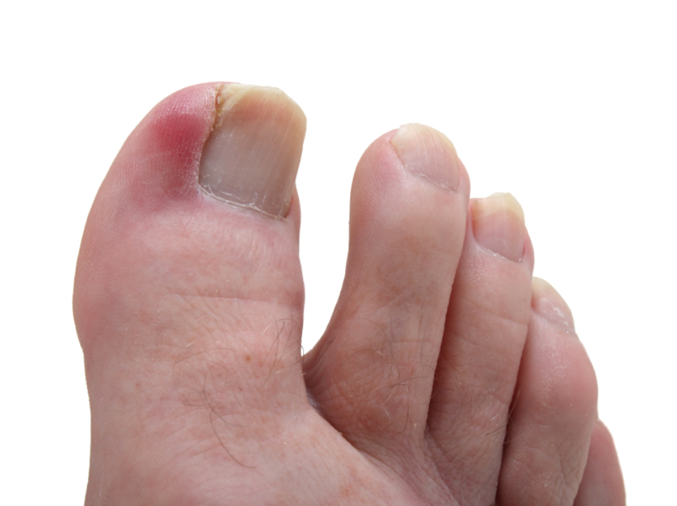
This infection, typically caused by Staphylococcus or Streptococcus, attacks the soft tissue borders surrounding the nail. Within 24 to 48 hours of an injury like biting a hangnail, the skin turns bright red, tight, shiny, and swollen. The digit will feel distinctly hot to the touch and will throb in sync with the patient’s pulse. Even the light pressure of a bedsheet can cause severe pain. As the infection progresses, a pocket of yellow or white pus will typically form beneath the cuticle or along the lateral nail fold, producing a foul odor.
Green Nail Syndrome
Green Nail Syndrome, or chloronychia, is caused by Pseudomonas aeruginosa. This bacterium thrives in wet environments and regularly colonizes nails that have already detached from their bed. As the bacteria multiply in the damp space beneath the loose nail, they produce unique pigments called pyocyanin and pyoverdine. These pigments stain the underside of the nail plate a striking shade of fluorescent green, dark forest green, or blackish-blue. Unlike paronychia, Green Nail Syndrome may not always cause severe throbbing pain if it is confined to a dead space, but it spreads rapidly across the surface and has a distinct, sickly-sweet odor.

4. Medical Diagnostic Protocols
Self-diagnosis based on visual appearance alone can lead to errors. A professional podiatric or dermatological evaluation relies on specific objective laboratory and clinical tests.
Fungal Diagnostic Assays
Clinicians use Potassium Hydroxide (KOH Prep) testing by scraping debris from beneath the nail plate, mixing it with a KOH solution, and examining it under a microscope. The solution dissolves the human keratin, leaving the rigid fungal hyphae walls clearly visible.
Alternatively, a fungal culture involves placing nail trimmings in a growth medium for several weeks to identify the exact species of fungus, though it takes a long time to get results. Polymerase Chain Reaction (PCR) testing is the modern gold standard, identifying the DNA of specific fungal pathogens within days for a rapid and precise diagnosis.
Bacterial Diagnostic Assays
If a fluid pocket or pus is present, the clinician will perform a wound swab and culture, expressing a small amount of fluid onto a sterile swab to isolate the specific bacterial strain on agar plates.
Gram staining is a quick laboratory technique that categorizes the bacteria as Gram-positive (like Staphylococcus) or Gram-negative (like Pseudomonas), allowing the physician to select an empirical antibiotic immediately while awaiting full culture results.
5. Treatment Modalities for Onychomycosis
Because fungal spores are buried within a dense, non-vascular structure, delivering treatment to the root of the infection requires specialized approaches.
Topical Antifungal Lacquers
Topical solutions (such as ciclopirox, efinaconazole, or tavaborole) are painted directly onto the clean nail plate daily. This approach is extremely safe with virtually no systemic side effects or drug interactions. However, the dense keratin of the nail plate acts as a highly effective barrier, making it very difficult for the medication to reach the nail bed. Clear rates can be modest, and the treatment demands months of strict compliance.
Oral Antifungal Medications
Systemic medications (like terbinafine or itraconazole) are taken orally, traveling through the bloodstream to enter the nail matrix from the inside out, infusing the newly growing nail with antifungal properties. While this method offers significantly higher success and clearance rates than standard surface topicals, these medications pass through the liver and can cause hepatotoxicity. Patients must undergo routine liver enzyme blood tests. They also carry a high risk of drug interactions, making them unsuitable for many older adults or individuals with pre-existing conditions.
Laser Nail Therapy (Light-Based Destruction)
Laser therapy addresses the core limitation of topicals (penetration) and oral medications (liver toxicity), completely avoiding systemic medications. Podiatric lasers utilize specific light wavelengths, such as 1064nm Nd:YAG lasers. This light passes through the clear and damaged structures of the nail plate without being absorbed by normal human tissue. However, when the light hits the fungal cells and their pigments, it converts into targeted thermal energy.
The intense, rapid pulses of heat vaporize the fungal hyphae and spores trapped within the keratin layers. Because the pulse intervals are highly calibrated, the underlying dermis has time to dissipate heat, preventing burns to the nail bed while maintaining a lethal temperature for the fungal colony. The laser does not instantly clear the physical discoloration. Instead, it sterilizes the nail unit. Healing is visible as the matrix produces healthy, uninfected keratin, slowly pushing the old, damaged nail out over the course of 6 to 12 months.
6. Treatment Modalities for Bacterial Nail Infections
Because bacterial infections escalate rapidly and occur in vascular tissue, they require immediate intervention to stop the spread of infection. Laser nail therapies designed for fungus are not appropriate for active bacterial nail infections. Instead, medical protocols focus on drainage and chemical eradication.
Decompression and Clinical Drainage
If a patient presents with an acute paronychia containing a visible abscess of pus, a medical professional must drain the fluid pocket. Under sterile conditions, a clinician gently separates the nail fold from the plate or uses a fine-gauge needle to puncture the abscess. Relieving the pressure provides immediate pain relief and prevents the pocket from cutting off blood supply to the local tissue matrix.
Targeted Antibiotic Therapy
For localized, mild infections, prescription-strength topical antibiotic ointments like mupirocin are applied to the tissue border. If the infection shows signs of tracking down the digit or is accompanied by red streaking, oral antibiotics (such as cephalexin or doxycycline) are prescribed to eliminate the bacteria systemically.
Environmental Alteration (For Green Nail Syndrome)
Because Pseudomonas is highly sensitive to acidic environments, mild or superficial cases of Green Nail Syndrome are often treated by altering the local environment. This involves regularly soaking the nail in a dilute solution of acetic acid (vinegar) or applying specialized topical solutions designed to lower the pH of the nail bed, making it impossible for the bacteria to survive.
7. Can Fungi and Bacteria Co-Infect the Same Nail?
Yes. In clinical dermatology, this is known as a microbial co-infection or secondary bacterial invasion.
The process typically follows a predictable sequence beginning with a primary case of chronic onychomycosis. As the fungus eats away at the structural anchors of the nail bed, it causes the nail plate to pull upward and separate, a state called onycholysis. This separation creates a dark, hollow, stagnant micro-pocket beneath the nail. When the patient showers or swims, water gets trapped inside this newly formed dead space. Pseudomonas bacteria migrate into the moisture pocket, establishing a colony right alongside the pre-existing fungus.
When a co-infection occurs, the nail may turn a muddy green-brown and crumble simultaneously. In these scenarios, a multi-phased treatment approach is necessary. The clinician must first eliminate the active bacterial population and dry out the space before utilizing laser therapy or topicals to clear the deeper fungal infection.
8. Long-Term Prevention and Maintenance
Regardless of which pathogen you have previously encountered, maintaining proper hand and foot hygiene is the best defense against reinfection.
Moisture Management
Always dry the spaces between your toes with a clean towel immediately after bathing. Wear moisture-wicking socks made of synthetic blends, merino wool, or bamboo, and avoid pure cotton socks, which trap dampness against the skin. Additionally, avoid wearing the same pair of shoes two days in a row, giving footwear at least 24 hours to air out and completely dry inside.
Structural Protection
Cut nails straight across rather than rounding out the corners. Rounding the edges increases the risk of developing an ingrown nail, which tears the skin barrier and invites bacteria. Never use unsterilized nail clippers, files, or cuticle pushers. If you visit professional salons, ensure they use hospital-grade autoclaves to clean their instruments. Finally, always wear sandals, flip-flops, or water shoes when walking in high-risk public spaces like gym locker rooms, public showers, or pool decks.
Summary: When to Seek Professional Care
Understanding the differences between these two conditions helps you identify when a nail issue requires urgent care versus routine management.
If you notice a nail slowly changing color, getting thicker, or becoming brittle over several months, you are likely dealing with onychomycosis. This is a chronic issue that can be discussed during a standard clinical visit to explore clearance options like laser therapy or targeted medications.
However, if you experience sudden redness, swelling that spreads down your digit, hot skin, or throbbing pain, you may have an acute bacterial infection. These issues may require prompt medical attention from a physician or podiatrist for next steps on protecting the nail matrix and ensure the infection is contained.